Kidney Blood Vessels Labeled : Kidney blood vessels, confocal micrograph - Stock Image ... / The smallest arteries give rise to afferent arterioles in the renal.. Blood vessels in parenchy… category: There is a printable worksheet available for download here so you can take the quiz with pen and paper. The function of kidney and blood in clearing wastes is evident from the fact weight of kidney is less than 1% (one percent) of total body weight while receiving 20. Our engaging videos, interactive if you're learning about kidney anatomy, you might like our urinary system quizzes and labeled diagrams! The blood vessels are the components of the circulatory system that transport blood throughout the human body.
The difference in the structural characteristics of arteries, capillaries and veins is attributable to their respective functions. Blood vessels of the kidney. These vessels transport blood corpuscles, nutrients, and oxygen to the tissues of the body. Blood vessels are vital for the body and play a key role in diabetes helping to transport glucose and insulin. Blood vessels in parenchy… category:
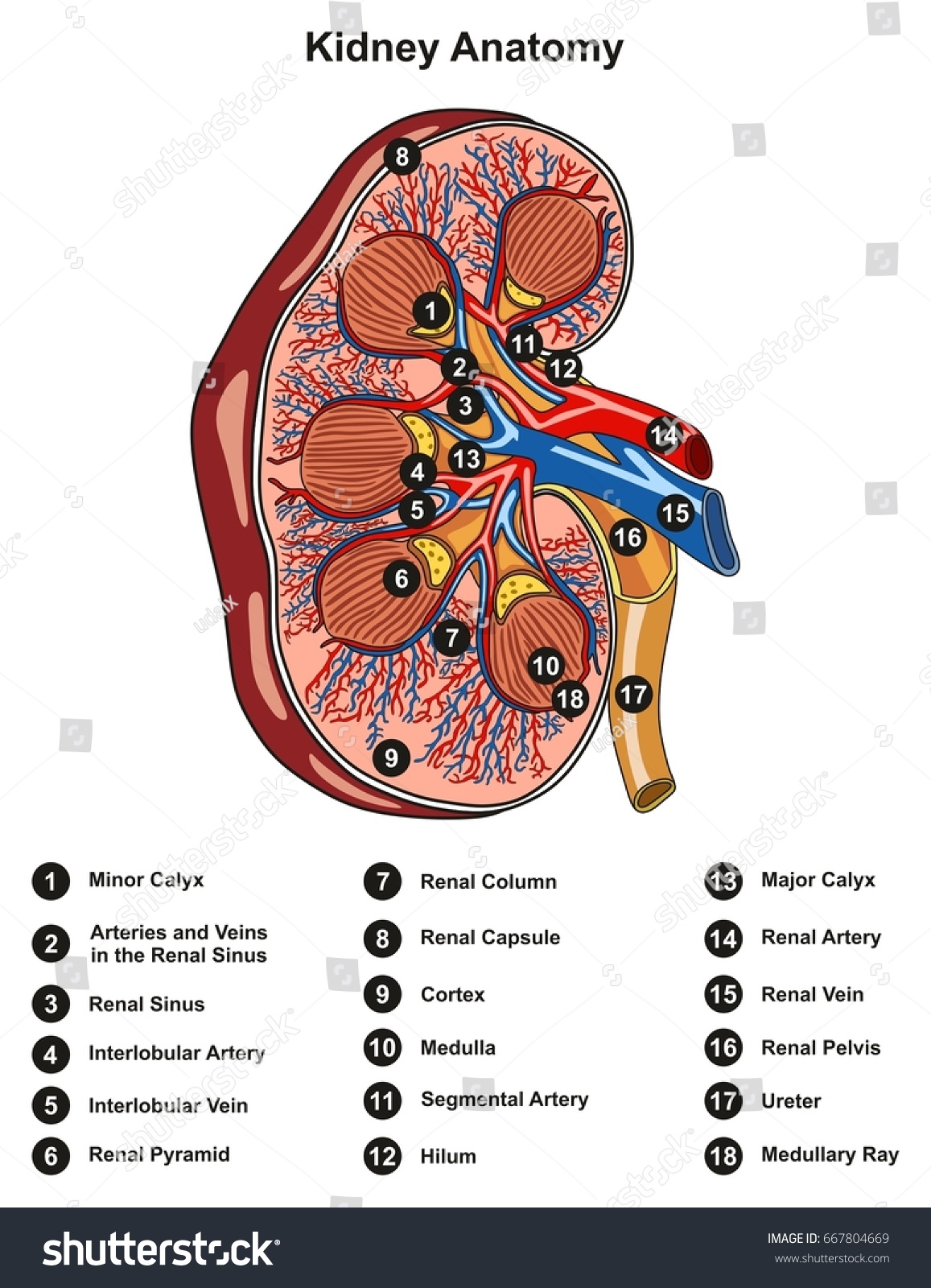
Kidney - Blood supply and Innervation | Kenhub from thumbor.kenhub.com The pathway of blood flow through the kidney is an essential part of the process of urine formation. Our kidneys regulate the water concentration in our blood and excrete toxic waste. Interlobar vein interlobular artery renal vein segmental artery arcuate vein renal artery interlobar artery interlobular vein arcuate artery reset zoom. Blood vessels are the branches of the circulatory system that carries blood in the human body. Labeled kidney anatomy cross section infographic diagram including all parts renal pelvis calyx medulla cortex ureter artery and vein supply blood vessels for medical science education and health care. The endothelial cells, which line the luminal surface of the blood vessels, have flattened to ovoid nuclei. Depression in the kidney is called hilus, from where blood vessel leaves and enter the kidney. The blood supply to the kidneys originates from the paired renal arteries, which branch into segmental arteriesat the renal hilum.
This article covers the blood supply, innervation, lymphatic drainage of the kidneys and related neurovascular supply of the kidney:
The endothelial cells, which line the luminal surface of the blood vessels, have flattened to ovoid nuclei. Efferent arterioles that are located above the corticomedullary border travel downward into that depends on which what kind of blood vessel you cut, and how much of it is damaged. They also take waste and carbon dioxide away from the tissues. Therefore, there is considerable interest in the haemodynamic and molecular mechanisms that may be responsible for alterations in the vascular. Because the kidney filters blood, its network of blood vessels is an important component of its structure and function. Want to learn more about it? Kidney section, nephrons, blood vessels and renal corpuscle. It carries the urea loaded blood into the glomerulus of the kidney. 2,731 blood vessels labeling machine products are offered for sale by suppliers on alibaba.com, of which labeling machines accounts for 5. The function of kidney and blood in clearing wastes is evident from the fact weight of kidney is less than 1% (one percent) of total body weight while receiving 20. The blood supply to the kidneys originates from the paired renal arteries, which branch into segmental arteriesat the renal hilum. Locate the renal capsule, the renal cortex (which appears somewhat granular and may. The basic physiology of a nephron within a kidney :
Labeled kidney anatomy cross section infographic diagram including all parts renal pelvis calyx medulla cortex ureter artery and vein supply blood vessels for medical science education and health care. Want to learn more about it? It carries the urea loaded blood into the glomerulus of the kidney. The pathway of blood flow through the kidney is an essential part of the process of urine formation. Galectin 3 is a member of the multifunctional galectin family, which is ubiquitously expressed in the heart, the kidney, blood vessels, and macrophages and plays a role in tissue fibrosis, immunity, and the inflammatory response.
Kidney Anatomy Cross Section Infographic Diagram Stock ... from image.shutterstock.com Milhares de fotos novas de alta qualidade são adicionadas todos os dias. The blood supply to the kidneys originates from the paired renal arteries, which branch into segmental arteriesat the renal hilum. Labeled kidney anatomy cross section infographic diagram including all parts renal pelvis calyx medulla cortex ureter artery and vein supply blood vessels for medical science education and health care. They regulate the water content in the blood. A part of the blood from the left ventricle is transferred to the kidneys through the renal artery for ultra purification. Correctly label the blood vessels of the kidney. Procedure b the renal blood vessels and nephrons 1. Vessels labeled diagram, blood vessels labeling exercises, cat blood vessels labeled, human anatomy blood vessels, human blood.
Our engaging videos, interactive if you're learning about kidney anatomy, you might like our urinary system quizzes and labeled diagrams!
Blood vessels play a key role in the progression of renal damage in aging, with reductions in glomerular filtration rate and renal blood flow. Transfection of isolated blood vessel endothelial cells to overexpress the lymphoendothelial. Kidney section, nephrons, blood vessels and renal corpuscle. The blood supply to the kidneys originates from the paired renal arteries, which branch into segmental arteriesat the renal hilum. Our engaging videos, interactive if you're learning about kidney anatomy, you might like our urinary system quizzes and labeled diagrams! Label figure complete part b of the laboratory report. Therefore, there is considerable interest in the haemodynamic and molecular mechanisms that may be responsible for alterations in the vascular. Identify the blood vessels (red/blue) pointed to by the arrow (use one line on answer sheet). When they fail to work properly, dialysis treatment or a transplant is the kidneys are located in the back of the abdomen and have two important functions in the body: They also take waste and carbon dioxide away from the tissues. • identification of blood vessels as arteries, capillaries or veins from the structure of their walls. Galectin 3 is a member of the multifunctional galectin family, which is ubiquitously expressed in the heart, the kidney, blood vessels, and macrophages and plays a role in tissue fibrosis, immunity, and the inflammatory response. Want to learn more about it?
A blood vessel that is part of a kidney automatically generated definition. Blood vessel disorders of the kidneys have a number of causes, including blockages in the renal arteries or veins, inflammation of blood vessels (vasculitis), injury to the kidneys or blood vessels, and other disorders. The blood vessels are the components of the circulatory system that transport blood throughout the human body. These vessels transport blood corpuscles, nutrients, and oxygen to the tissues of the body. Arteries are the blood vessels that carry blood away from the heart.
Vessel Lab from faculty.collin.edu These vessels transport blood corpuscles, nutrients, and oxygen to the tissues of the body. Correctly label the blood vessels of the kidney. Blood from the abdominal aorta enters the renal artery, which branches extensively within the kidney into smaller arteries (see fig. The blood supply to the kidneys originates from the paired renal arteries, which branch into segmental arteriesat the renal hilum. We'll assume for the purposes of this answer that the. The endothelial cells, which line the luminal surface of the blood vessels, have flattened to ovoid nuclei. The basic physiology of a nephron within a kidney : Therefore, there is considerable interest in the haemodynamic and molecular mechanisms that may be responsible for alterations in the vascular.
Blood vessels are the branches of the circulatory system that carries blood in the human body.
4.which blood vessel will have the high amount of glucose and amino acld after a meal? Interlobar vein interlobular artery renal vein segmental artery arcuate vein renal artery interlobar artery interlobular vein arcuate artery reset zoom. Identify the layer of the kidney indicated by the curly bracket. They regulate the water content in the blood. Arteries are the blood vessels that carry blood away from the heart. Our kidneys regulate the water concentration in our blood and excrete toxic waste. Blood vessels of the kidney. The blood vessels are the components of the circulatory system that transport blood throughout the human body. Because the kidney filters blood, its network of blood vessels is an important component of its structure and function. Locate the renal capsule, the renal cortex (which appears somewhat granular and may. Milhares de fotos novas de alta qualidade são adicionadas todos os dias. Want to learn more about it? Identify this structure (tube) 13.
⇒ click on the diagram to show / hide labels blood vessels labeled. The blood vessels are the components of the circulatory system that transport blood throughout the human body.


0 Komentar